.jpg)
.jpg)
Livestock: Cow
Breeds of Dairy Cattle
Indigenous Cows
?The Indian milch breeds are Sahiwal, Red Sindhi, Gir.
?The milk production is on an average 1600 kg per lactation.
Sahiwal
Sahiwal
?Originates from Montgomery district of Pakistan
?More population of these breeds are found in Punjab, Haryana, U.P, Delhi, Bihar and M.P.
?Milk yield ranges from (1350 kg to 2100 kg).
?Age at first calving (32-36 months )
Red Sindhi
.jpg) Red Shindhi
Red Shindhi
?Belongs to Kohistan, Sindh province in Pakistan.
?It is one of the most distinctive cattle breeds of India.
?Mainly available in Punjab, Haryana, Karnataka, Tamil Nadu, Kerala and Orissa.
?Milk yield varies from (1700 kg -3400 kg per lactation).
Gir
?Mainly found in Gir forest areas of South Kathiawar.
?Milk yield varies from (900 -1600 kg in lactation)
Exotic Breeds of Dairy Cow
Jersey
.jpg) Jersey
Jersey
?Developed from the island of jersey, as France.
?The Jerseys coat colour varies from a very light gray to a very dark fawn or a shade that is almost black.
?Age at first calving : 26-30 months.
?Milk yield ?5000-8000 kg.
?Jersey yield 20 litres/day whereas, crossbred jersey cow gives 8-10 litres/day.
?Well developed to the climatic condition of India especially hot and humid areas.
Holstein Friesian
.jpg) Holstein Friesian
Holstein Friesian
?This breed is originated in Holland.
?Holsteins are large capacious animals with colour patterns of black and white or red and white.
?Milk yield - 7200-9000 kg the best diary breed among exotic cattle regarding milk yield.
?On an average it gives 25 liter of milk per day, whereas a cross breed H.F. cow gives 10 - 15 liter per day.
?It can perform well in coastal and delta areas.
Cross-breed cattle
The crossbreeds are having exotic inheritance from Jersey, Brown Swiss or Holstein Friesian or a combination of these different breeds. Jersey breed is known for the milk fat percent and Holstein for the high quantity of milk.
Proper selection is the first and the most important step to be adopted in dairying. Records are the basis of selection and hence proper identification of animals and record keeping is essential. Cross-breed animals with exotic inheritance of about 50 percent are preferable. This preference is based on comparison of the performance of the animals with different percentage of exotic inheritance. Fifty percent of the native germplasm is helpful to retain the adaptability, heat tolerance and disease resistance traits of local animals, in cross breeds. The utilization of the Zebu (Sahiwal) germplasm in the formation of breeds like Australian Friesian Sahiwal (50% of Holstein and 50% Sahiwal) and its international recognition as a breed for the tropics is an example.
Maintaining animals sustainable to the situation is the best policy. Bringing animals from different agro-climatic conditions causes problems due to non-adjustment in many cases. In case, purchase becomes absolutely essential it should be from similar environmental conditions as far as possible.
General selection procedures for dairy breeds
Selecting a calf in calf show, a cow in cattle show by judging is an art. A dairy farmer should build up his own herd by breeding his own herd. Following guidelines will be useful for selection of a diary cow.
?whenever an animal is purchased from a cattle fair, it should be selected based upon its breed characters and milk producing ability.
?History sheet or pedigree sheet which are generally maintained in organized farms reveals the complete history of animal.
?The maximum yields by dairy cows are noticed during the first five lactations. So generally selection should be carried out during First or Second lactation and that too are month after calving.
?There successive complete milking has to be done and an average of it will give a fair idea regarding production by a particular animal.
?A cow should allow anybody to milk, and should be docile.
?It is better to purchase the animals during the months of October and November.
?Maximum yield is noticed till 90 days after calving.
Breed characteristics of high yielding dairy cows
?Attractive individuality with feminity, vigour, harmonious blending of all parts, impressive style and carriage ?Animal should have wedge shaped appearance of the body.
?It should have bright eyes with lean neck.
?The udder should be well attached to the abdomen.
?The skin of the udder should have a good network of blood vessels.
?All four quarters of the udder should be well demarcated with well placed teats.
Selecting breeds for Commercial Dairy Farm - Suggestions
?Under Indian condition a commercial dairy farm should consist of minimum 20 animals (10 cows, 10 buffaloes) this strength can easily go up to 100 animals in proportion of 50:50 or 40:60. After this however, you need to review your strength and market potential before you chose to go for expansion.
?Middle class health-conscious Indian families prefer low fat milk for consumption as liquid milk. It is always better to go for a commercial farm of mixed type. (Cross breed, cows and buffaloes kept in separate rows under one shed).
?Conduct a through study of the immediate market where you are planning to market your milk You can mix milk from both type of animals and sold as per need of the market. Hotels and some general customers (can be around 30%) prefer pure buffalo milk. Hospitals, sanitariums prefer cow's milk.
Selection of cow breeds for commercial farm
?Good quality cows are available in the market and it cost around Rs.2500 to Rs.3000 per liter of milk production per day. (E.g. Cost of a cow producing 10 liter of Milk per day will be between Rs.25000 to Rs.30,000).
?If proper care is given, cows breed regularly giving one calf every 13-14 month interval.
?They are more docile and can be handled easily. Good milk yielding cross breeds (Holstein and Jersey crosses) has well adapted to Indian climate.
?The fat percentage of cow's milk varies from 3-5.5% and is lower then Buffaloes.An efficient management of cattle will be incomplete without a well planned and adequate housing of cattle. Improper planning of animal housing leads to additional labour charges. The housing should have proper sanitation, durability, arrangements for the production of clean milk under convenient and economic conditions.
Location of Dairy Buildings
The points which should be considered before the creation of dairy buildings animal houses are as follows.
Topography and drainage
The houses should be well raised / elevated for the surrounding ground to offer a good slope for rainfall and more drainage of dairy wastes to avoid stagnation and for the spread of diseases. A levelled area requires less site preparation and thus lesser cost of building. Low lands and depressions should be avoided.
Exposure to the sun and protection from wind
A dairy building should be located to a maximum exposure to the sun in the north and minimum exposure in the south and protect from prevailing strong wind currents whether hot or cold. Buildings should be placed such that direct sunlight can reach the platforms, gutters and mangers in the cattle shed. Its better to have, the long axis of the dairy barns set in the north-south direction to have maximum benefit of the sun.
Water supply
Abundant supply of fresh, clean and soft water should be available.
Surroundings
Narrow gates, high manger curbs, loose hinges, protruding nails, smooth finished floor in the areas where the cows move should be eliminated.
Labour
Honest, economic and regular supply of labour should be available.
Marketing
Dairy buildings should be in those areas where selling of dairy products can be done profitably and regularly. Owner should be in a position to satisfy the needs of the farm within no time and at reasonable price.
Facilities, labour, food
Cattle yards should be situated in relation to feed storages, hay stacks, silo and manure pits as to effect the most efficient utilization of labour. Sufficient space per cow and well-arranged feeding mangers and resting contribute not only to greater milk yield of cows and make the work of the operator easier also minimizes feed expenses.
Cattle Shed
The entire shed should be surrounded by a boundary wall of 5" height from three side and manger etc., on one side. The feeding area should be provided with 2 to 2 ½ feet of manger space per cow. All along the manger, there shall be 10" wide water trough to provide clean, even, available drinking water. The water trough constructed can minimize the loss of fodders during feeding. Near the manger, under the roofed house 5" wide floor should be paved with bricks having a little slope. Beyond that, there should be open unpaved area (40'X35') surrounded by 5" wall with one gate. It is preferable that animals face north when they are eating fodder under the shade. During cold wind in winter the animals will automatically lie down to have the protection from the walls.
Cow sheds can be arranged in a single row if the numbers of cows are small. In double row housing, the stable should be so arranged that the cows face out (tails to tail system) or face in (head to head system) as preferred.
Floor
The inside floor of the barn should be of some impervious material which can be easily kept clean and dry and is not slippery. Grooved cement concrete floor is still better. The surface of the cowshed should be laid with a gradient of 1" to 1 14" from manger to excreta channel. An overall floor space of 65 to 70 sq.ft. Per adult cow should be satisfactory.
.jpg)
.jpg)
Wall
The inside of the walls should have a smooth hard finish of cement, which will not allow any lodgment of dust and moisture. Corners should be round. The open space in between supporting pillars will serve for light and air circulation.
Roof
Roof of the barn may be of asbestos sheet or tiles. However, iron sheets with aluminum painted tops to reflect sunrays and bottoms provided with wooden insulated ceilings can also achieve the objective. A height of 8 feet at the sides and 15 feet at the ridge will be sufficient to give the necessary air space to the cows. An adult cow requires at least about 800 cubic feet of air space under tropical conditions.
Manger
Cement concrete continuous manger with removable partitions is the best from the point of view of durability and cleanliness. A height of 1 '-4" for a high front manger and 6" to 9" for a low front manger is considered sufficient. Low front mangers are more comfortable for cattle but high front mangers prevent feed wastage. The height at the back of the manger should be kept at 2'-6" to 3". An overall width of 2' to 2 1/2' is sufficient for a good manger.
Labour
Honest, economic and regular supply of labour should be available.
Alleys
The central walk should have a width of 5'-6' exclusive of gutters when cows face out, and 4'-5' when they face in. The feed alley, in case of a face out system should be 4' wide, and the central walk should show a slope of 1" from the center towards the two gutters running parallel to each other, thus forming a crown at the center.
Manure Gutter
The manure gutter should be wide enough to hold all dung without getting blocked, and be easy to clean. Suitable dimensions are 2" width with a cross-fall of 1?away from standing. The gutter should have a gradient of 1" for every 10' length. This will permit a free flow of liquid excreta.
Door
The doors of a single range cowshed should be 5" wide with a height of 7', and for double row shed the width should not be less than 8" to 9'. All doors of the barn should lie flat against the external wall when fully open.
Sheds for Young Stocks
Calves should never be accommodated with adults in the cow shed. The calf house must have provision for daylight ventilation and proper drainage. Damp and ill-drained floors cause respiratory trouble in calves to which they are susceptible. For an efficient management and housing, the young stock should be divided into three groups, viz., young calves aged to one year bull calves, female calves. Each group should be sheltered in a separate calf house or calf shed. As far as possible the shed for the young calves should be quite close to the cow shed.
Care and management of calf
?Identification of calves can be done by tattooing of ear at birth, and branding after one year.
?Dehorn the calf within 7-10 days after birth with red hot iron or caustic potash stick or electrical method.
?Deworm at 30 days interval.
?Fresh water should be given age of 2 -3 weeks onwards.
?House the calves in individual calf pens for 3 months afterwards in groups. After six months males and females calves should be housed separately.
?Weigh the calves at weekly interval up to 6 months arid at monthly interval afterwards to know the growth rate.
?Mortality in calves is more in first month due to pneumonia, diarrhoea (calf scours) and worms. House them under warm, clean condition to avoid above problems.
?Extra teats beyond 4 should be removed at 1-2 months of age.
?Males should be castrated at 8-9 weeks of age.
?Keep the body clean and dry to avoid fungal infection.
?Mineral-blocks should be provided, so that the calves lick and no changes for mineral deficiency.
Care and management of Heifer
Better care and management of heifer will give high quality replacement stock to the dairy farm.
During the early stage relatively more protein than energy is needed. Most heifers grow well if excellent hay is given. The amount of growth depends upon the quality of forage fed. Feed the heifer sufficiently to produce normal growth.
?The heifers should be provided with a dry shelter free from drafts. A loose housing system with a shelter open to one side is sufficient.
?Breeding under sized animals is never profitable. Small heifers are more likely to have difficulty in calving.
?Place the heifer in a separate shed about 6-8 weeks before she is due to calve.
?Feed 2 - 3 kg of concentrate daily and all other green.
?Animals lagging behind below the required standards should be removed from the herd.
?For the heifer the calving is first time and it may have difficulty in calving. So take extra care during calving.
Care and management of Milch Animal
To get high milk during any lactation, the milch animal should be properly fed along a proper care and management practices.
?Provide green succulent forage together with leguminous hay or straw to the extent of animal can consume, so that all its maintenance requirements are met with through forage only. Extra concentrate at the rate of 1 kg for every 2 to 2.5 liters of milk should be provided. Salt and mineral supplements should be given to maintain the lactation.
?Never frighten or excite the animals.
?Concentrate mix is fed before or during milking, where as roughages after milking. This practice will avoid dust in the shed.
?Water should be provided adlibitum.
?Regularity in milking is essential. Increase of milk in the udder will reduce further secretion of milk. Milking thrice is better than twice since 10 - 15 % more milk can be produced.
?Milking should be done with whole hand.
?Cows should be trained to let down milk without calf suckling. This will held to wean the calves early.
?Loose housing with shelter during hot part of the day should be provided.
?The animals will get maximum exercise in loose housing system.
?Grooming of the cows and washing of the buffaloes before milking help in clean milk production.
?Daily brushing will remove loose hair an dirt from the coat.
?Grooming will also keep the animal hide pliable.
?Wallowing of buffaloes or water spraying on their bodies will keep the buffaloes comfortable especially in summer.
?Provide at least 60 days dry period between calving. If the dry period is not sufficient, the milk yield is subsequent lactation will be reduced.
?Vaccinate the cows- against important diseases.
?Every animal should be numbered and particulars pertaining to milk, fat %, feed taken, breeding, drying and calving dates should be recorded.
?Check for mastitis regularly.
Animal Feeding
Good Practice
?Timely provision of feed to meet livestock nutritional requirements.
?Always give the animals clean and healthy feed.
?Input must be adequate in both nutritional and economic terms. Feeding cows according to the stage of lactation.
?Make sure adequate water available after milking.
?Cows need clean potable water and free access to it.
Bad Practice
?Insufficient quantity of feed supplied.
?Use of poor quality feed stuffs, Inadequate to meet the animals requirement, Feeding unbalanced rations leads to wastage and poor performance.
?Feeding fodder that has been recently sprayed with pesticide.
?Using dirty water for drinking.
Environment
Good Practice
?Cleaning away waste every day.
?Separating medicine and chemical containers from other organic waste.
?Use the manure for land improvement.
?Keep the area clean and safe for animals.
Bad Practice
?Wet unventilated dirty environment.
?Burning rubbish in the vicinity of the cows.
?Excessive use of chemicals, fertilizers and pesticides, contaminates the soil and ground water.
Milking Hygiene
Good Practice
?Washing hands with soap and water before milking each cow. Washing of hands thoroughly between finishing milking one cow and beginning the milking of the next.
?Washing the udder and each teat vigorously with soap and water and dry them with a clean cloth.
?Direct the first milk outside the milking bucket into separate container and throw away.
?Have a clean, dry, floor preferably of rough surfaced concrete without sharp points for the milking area.
?Keep calves where cows can see them during milking.
?Use clean containers for milking and before re-using the milk container: rinse it, scrub it with warm water and detergent or soap, rinse it again leave it to air-dry.
?After milking, cover the milk to avoid contamination and place in a clean and cool area.
?Keep the area clean and safe for animals.
Bad Practice
?Using Milk from sick cows can transmit diseases to humans.
?Using unclean plastic containers.
?leaving milk uncovered.
?Keeping the milk in the sun or outdoors.
Method of Milking
Hand milking and machine milking are the two methods.
Many milkers during milking tend to bend their thumb against the teat. The method is known as knuckling which causes injury to teat tissues. Thus milking should always be done with full hand unless the teats are too small or towards the completion of milking. The first few strips of milk from each quarter should not be mixed with the rest of the milk as the former contains highest number of bacteria.
Machine Milking
Modern milking machines are capable of milking cows quickly and efficiently, without injuring the udder. The milking machine performs two basic functions.
?It opens the streak canal through the use of a partial vacuum, allowing the milk to flow out of the teat cistern through a line to a receiving container.
?Massages the teat, preventing congestion of blood and lymph in the teat.
Advantages
Easy to operate, costs low, saves time as it milks 1.5 litre to 2 litres per minute. It is also very hygienic and energy-conserving as electricity is not required. All the milk from the udder can be removed. The machine is also easily adaptable and gives a suckling feeling to the cow and avoids pain in the udder as well as leakage of milk.
Clean Milk Production :
Milk containing dirt, dust, foreign materials high bacterial count and with off, flavour is called contaminated milk.
Milk is contaminated by various sources :
Udder
Unsanitary conditions of milking barns and bedding of the animal causes bacterial growth. Such bacteria may enter in to the udder through teat canal, which causes infection the udder like mastitis resulting contamination of milk. The fore milk may be discarded as it contains high bacterial count. Complete milking should be done. Incomplete milking may lead to infection of the udder.
Exterior of cow's body
Bacteria present on the animal body may enter in to the milk at the time of milking. Maintenance of clean skin, washing flank and udder with clean damp cloth before milk reduces the contamination from this source.
Milking barns
Provided with good ventilation and neat flooring.
Dry feeds or forage should be fed after milking.
Milker
Dirty hands and clothing of the milker may be the source of contamination causing spread of bacterial diseases to the consumer through milk. Persons suffering from diseases like T.B, Typhoid fever, diphthiria may not be employed for milking. Dirty habits like smoking, drinking should be avoided.
Utensils
Clean sanitized, smooth copper free and dry utensils may be used for handling milk.
Detergents and disinfectants
Detergents increase the 'wetting' potential over the surfaces to be cleaned, displace milk deposits, dissolve milk protein, emulsify the fat and aid the removal of dirt. Detergent effectiveness is usually increased with increasing water temperature, and by using the correct concentration and time of application.
Detergents contain inorganic alkalis (eg. sodium carbonate and silicates and tri-sodium phosphate), surface-active agents (or wetting agents), sequestering (water-softening) agents (eg. polyphosphates) and acids for de-scaling. An inexpensive mixture can be made to give a concentration in solution of 0.25% sodium carbonate (washing soda) and 0.05% polyphosphate (Calgon). Disinfectants are required to destroy the bacteria remaining and subsequently multiplying on the cleaned surfaces. The alternatives are either heat applied as hot water or chemicals. Heat penetrates deposits and crevices and kills bacteria, providing that correct temperatures are maintained during the process of disinfection. When hot water alone is used, it is best to begin the routine with water at not less than 85°C, so that a temperature of at least 77°C can be maintained for at least 2 minutes.
If any concentrated detergent and/or disinfectant comes in contact with the skin or eyes the affected area should be washed immediately with copious amounts of clean water.
Milking premises
The milking premises should have a dairy or suitable place equipped with a piped hot and cold water supply, a wash trough, brushes, a work surface, storage racks and cupboards and, if necessary, a vacuum pipeline connection. In addition, it is advisable to have a dairy thermometer (0°C - 100°C), rubber gloves and goggles for use when handling chemicals.
Daily routines
Coolers, either the corrugated surface or the turbine in-can, can best be cleaned and disinfected manually and stored in the dairy to drain. Refrigerated bulk milk tanks can be cleaned either manually using cold or warm detergent/disinfectant solutions, or for the larger tanks, by automatic, programmed equipment. In either case, a cold water chlorinated (50 ppm) rinse proceeds and follows the washing solution. Foremilk cups can be a potent source of bacterial contamination and need to be cleaned and disinfected after each milking. They should then be stored in the dairy to drain.
It is important with any method of cleaning that the equipment is drained as soon as possible after washing for storage between milking. Bacteria will not multiply in dry conditions but water lodged in milking equipment will, in suitable temperatures, provide conditions for massive bacterial multiplication. Equipment with poor milk contact surfaces, crevices and large number of joints, remaining wet between milking in ambient temperatures above 20°C, should receive a disinfectant rinse (50 ppm available chlorine) before milking begins.
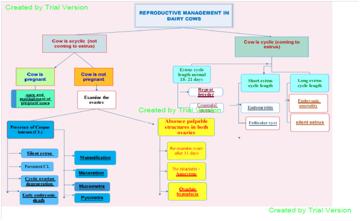
Reproductive management in dairy cows
Reduced fertility is one of the commonest reasons for vets being called onto dairy farms. In many cases cows presented to vets have obvious problems such as “whites?or ovarian cysts.
However, not every cow failing to hold to service has an obvious problem. A significant number (around 10 to 15%) of cows require four or more inseminations to get pregnant despite apparently cycling normally.
Most of these are indeed “normal?since, assuming no cows are culled after earlier services, then even in a herd with every cow becoming pregnant at a rate of 50% (a good rate for our dairy farms these days) there will be 12.5% of cows presented for a 4th service!
However, in some cases there is an underlying problem reducing the chance of the cow getting pregnant. These may be either cow factors that require ultrasound examination or even other more sophisticated tests e.g. oviduct patency, or the result of a wide range of external factors, such as poor management.
An obvious example is inefficient oestrous detection. On farm, without extensive testing, it is very difficult to distinguish between all these factors, as their only sign is a cow that is apparently normal but hasn’t got pregnant. Such cows get lumped together as having ‘repeat breeding syndrome?
CARE AND MANAGEMENT OF PREGNANT COW
-
Pregnant animals should be watched carefully, particularly during the last stages of pregnancy to avoid abortion due to fights or other physical trauma.
-
During early stages of pregnancy, there is no need of special feeding for heifers. The system of feeding and management recommended for heifers before breeding may continue. During last three months of pregnancy when fetal growth is very rapid, a special pregnancy allowance of about 1-2 Kg of concentrate should be offered.
-
Special care should be taken regarding mineral and vitamin deficiencies because they can have a serious adverse effect on the newborn calf. Feeding trace mineralized salt plus recommended amounts of calcium and phosphorus is usually sufficient to avoid these problems. Care must be taken that calcium and phosphorus should not be taken in excessive amounts.
-
During the last few weeks of pregnancy there is a tendency of prolapse of vagina which may be caused by constipation, mineral deficiency and debility. Balanced and laxative rations should be fed to maintain the normal tone of the reproductive tract.
-
Some time udder edema occurs before calving. This can be avoided by moderate exercise for a half an hour, two to three times per day. Massaging the udder for a few minutes is also helpful. Use of diuretics and prepartum milking may be helpful in severe cases.
-
Isolate the pregnant animal 8-10 days before the expected date of calving and keep it in a clean well bedded, dry and disinfected maternity pen. The animal should be watched closely as calving time approaches at least every two to three hours.
-
A good calving environment reduces the exposure of cows and newborn calves to infectious disease. A clean and comfortable area that provides cows with good footing minimizes the potential for injuries. Calving areas should be landscaped to allow for adequate drainage. Shade structures are recommended.
-
Calves are usually born without assistance. Any abnormality in their presentation requires immediate attention by a competent person to correct the position of the calf so that it can be delivered. Strict sanitation must be observed during assistance.
-
After removal of calf, milk animal it will help in removal of placenta. Placenta is normally expelled within 2 to 6 hours after calving. If placenta fails to be expelled with 12 hours it is considered retained placenta. In case of retained placenta veterinarian should be called for its removal.
-
After normal birth, the dam is alert and willing to eat and drink within one or two hours of calving. Warm water and some wheat bran should be offered to dam after calving. It is necessary to encourage the dairy animals to rise and to move to the manger for feeding after calving, especially on the day of calving and the first 2 days after calving.
-
The animal should be closely watched for health problems after calving. In addition to observing feed intake and milk production, rectal temperature and ketone levels should be monitored daily. Animals having health problems should be identified and treated accordingly, whereas healthy animals can join the general population 3 to 4 days postpartum.
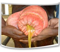
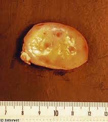
Cystic ovarian degeneration (COD) in cattle
Definition of COD
Traditionally cysts have been defined as anovulatory follicular structures (diameter, >25 mm) that persist for 10 or more days in the absence of a functional corpus luteum and are accompanied by abnormal oestrous behaviour (irregular oestrus intervals, nymphomania or anoestrus). However, recent data using ultrasonography indicate that follicles typically ovulate at 17 mm in diameter, so follicles that persist at 17mm or greater may be considered to be "cystic."
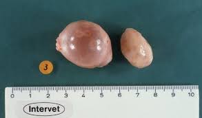
Nymphomanic behaviour Follicular cyst
Occurrence and consequences
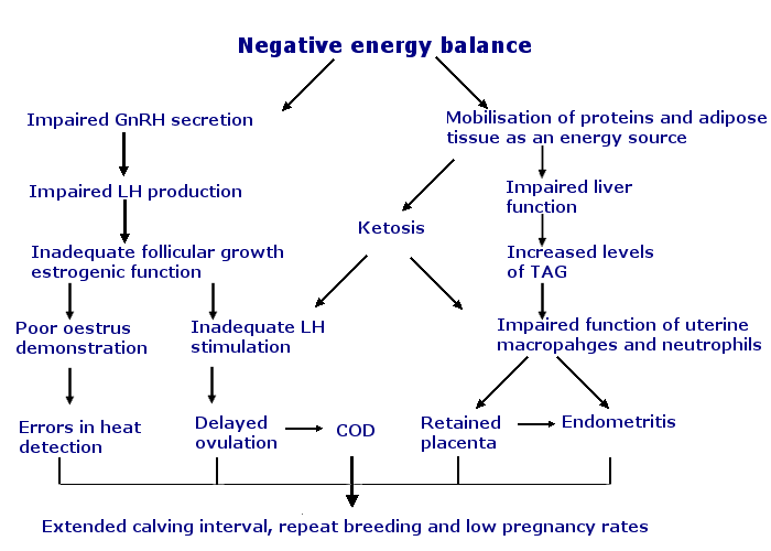
Cystic ovarian disease in dairy cattle occurs most frequently during the post partum period, 30 to 60 days after calving, when normal ovarian activity usually resumes.
The incidence of COD varies between 5 and 30 %. (Hooijer et al., 2001). Each incidence of ovarian follicular cysts increases the calving interval by 22-64 days.
Cause
Factors predisposing to cystic ovaries include metabolic diseases, negative energy balance, high productivity, retained placenta, dystocia, stress and genetics.
Follicular cysts result from failure of ovulation and luteinization. (Luteinization is the structural and biochemical changes that occur in a follicle after ovulation. Follicular cells that once produced estrogen change into luteal cells of the new corpus luteum (CL) that secrete progesterone. Follicular cysts are blister-like structures, flaccid to the touch.
Luteinized cysts apparently fail to ovulate, but some luteinization occurs. Because of the varying degree of luteinization, luteinized cysts are firmer to the touch than follicular cysts though not as solid as CL.
Cystic CL are CL with a fluid filled center. Several early researchers did not find cystic CL in pregnant cows and reasoned that cystic CL could not support pregnancy. However, Cornell University researchers reported that a CL producing only 100 µg of progesterone can support pregnancy. Therefore, a cystic CL could maintain pregnancy. In the absence of pregnancy, cystic CL regress and are considered nonpathological.
Symptoms
Abnormal estrous behavior patterns are the most noticeable sign of cystic ovarian disease.
This includes persistent oestrus, shortened oestrus intervals or failure to cycle (anoestrus). Anoestrus is by far the most common sign.
Persistent bulling behaviour, or nymphomania, is by far the least common clinical sign associated with cystic ovaries.
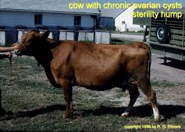
Sterility hump in cows with chronic cystic ovarian degeneration
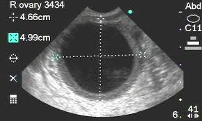
Ultra sound scanning of follicular cyst
Treatment
Early, prompt treatment is important as most cysts will not resolve on their own. Modern treatment of cystic ovaries in cattle addresses the underlying factors responsible for the development of the condition.
This could include correction of nutritional and metabolic disorders or hormonal treatment leading to induction of normal follicular turnover and resumption of cyclic ovarian activity.
Progesterone, prostaglandins and gonadotrophin-releasing hormones are the most commonly used products.
Prevention
The causes of ovarian cysts are not fully understood, which means there are no specific prevention regimes. However energy deficiency is a major factor and reducing the depth and length of the period of negative energy balance after calving, will significantly reduce the incidence of cystic ovaries.
http://www.thecattlesite.com/diseaseinfo/210/cystic-ovaries#sthash.NRUrX92P.dpuf
Early embryonic death
Embryonic mortality is generally defined as loss of the conceptus which occurs during the first 42 days of pregnancy, which is the period from conception to completion of differentiation when organ systems develop.
Approximately 30 percent of all embryos and fetuses will not survive to birth.
About 80 percent of this loss occurs before day 17, 10-15 percent between day 17 and 42 and 5 percent after day 42.
These losses to be much higher in "repeat breeders" those cows and heifers inseminated three or more times.
This has a significant economic impact on dairy herd profitability.
Because of the complex interactions among the various processes involved in establishment and maintenance of pregnancy, no single factor can be identified as the primary cause of embryonic death.
The following are some major factors contributing to this loss:
It is well documented that short term exposure to heat stress several days before and after insemination results in low conception rate or embryonic death. This is due to elevated temperature of the uterine environment. This has been a serious problem affecting reproductive performance this spring and summer.
Chromosomal abnormalities are known to be a cause of embryo mortality. One research summary estimated an average of 10 percent embryos had gross chromosomal abnormalities. Most of these embryos were lost before day 12. Unfortunately, this problem cannot be controlled through management.
Nutritional factors have been shown to contribute to low conception rates resulting from embryonic mortality. Research studies have shown that cows with severe change in body condition score (greater than 1 point loss on a scale of 1 to 5) during the first five weeks postpartum had an extended interval to first service and a very low conception rate compared to herdmates with only a minor or moderate change in body condition.
Whether this is due to an abnormal hormonal situation such as reduced progesterone secretion or ovulation of a defective oocyte has not been determined. Several reports showed that feeding excess crude protein, excess degradable intake protein or low levels of fermentable carbohydrate and the various combinations of these nutrients can cause low conception rates. Such situations can produce excessive levels of ammonia in the blood and uteri of cows.
Some researchers believe this could be toxic to the gametes and the developing embryo. The excess ammonia that is not utilized by the rumen microbes must be converted to urea at a significant energy cost which can further adversely impact conception rate. Mycotoxins and exposure to toxic substances and toxic plants have also been implicated as a cause of embryonic mortality.
Infectious agents can cause uterine infection or directly affect the embryo causing death. The major organisms adversely affecting reproduction are : Corynebacterium pyogenes, Campylobacter fetus (Vibriosis), Haemophilius somnus, Leptospirosis, Neospora and the viruses bovine virus diarrhea (BVD), infectious bovine rhinotracheitis (IBR) and to a lesser extent Ureaplasma and Mycoplasma.
Uterine defense mechanisms including the immune system are critical to maintaining embryonic development. For example, recent reports show that subclinical mastitis and other systemic infections can substantially increase the incidence of early embryo loss. Furthermore, nutritional deficiencies or an environmental challenge can alter uterine immune function. Currently this is a very active area of research.
Hormonal patterns or imbalances associated with embryonic mortality have been identified. The challenge for researchers is to develop synchronization programs and treatments that provide high progesterone, maintain low estrogen, and lead to a highly functional corpus luteum to maintain pregnancy. Dairy producers should continue to keep informed about new systems that will be developed to minimize embryonic loss. However, these systems will not correct problems associated with nutrition, disease, heat stress or postpartum problems that challenge the uterine defense mechanism.
There are management practices that can be implemented to minimize the incidence of embryonic mortality. These include: proper transition cow management and nutrition, effective vaccination, providing a clean environment, minimize heat stress and proper timing of insemination. There is abundant data to support the fact that between 5 percent and 30 percent of the cows inseminated are not in estrus when bred. Not only will this situation result in a low fertilization rate but most likely such cows are inseminated when the uterus is under the influence of progesterone and the immune system is not geared up to combat an infectious agent introduced during insemination.
Source: http://animalscience.psu.edu/news/2006/causes-of-embryonic-mortality-in-cattle
Mummification of the fetus
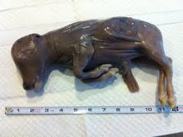
Mummification of the bovine fetus occurs in cattle of all ages. The condition usually affects single fetuses but may occasionally involve one or both fetuses in twin pregnancies. Bovine fetal mummification occurs in the third to eight month of gestation but most commonly the fourth, fifth and sixth months; if the condition is undiagnosed the mummified fetus will remain in the uterus for months beyond a normal gestation period.
Spontaneous abortion before or near the expected end if gestation are not uncommon. Conditions necessary for the occurrence of fetal mummification include the maintenance of the dead fetus within the uterus by the presence of a normal viable fetus or fetuses or the persistence of the corpus luteum.
Fetal mummification apparently does not occur in the first trimester of gestation because embryonic or fetal death prior to development of the fetal bones usually is followed by absorption or resorbtion of the fetal and placental tissues.
Fetal death the last month or six weeks of gestation will begin fetal mummification may be undiagnosed at parturition and called a stillbirth, especially in multiparous.
Causes of fetal mummification:
The causes of fetal death and mummification in cattle are often the same as causes for fetal and abortion. The cause of fetal mummification is often impossible to determine because the time of fetal death is not known and autolysis and mummification of the fetus and membranes makes determination of the causative agent difficult or impossible:
1- Genetic factors
2- Torsion of umbilical cord or compression of umbilical cord by its passing around a fetal extremity.
3- Infectious causes of fetal death such as V. fetus, molds, Leptospirosis?etc.
Diagnosis of F.M:-
Depending on:
1- Case history.
2- Rectal palpation.
As the fetus mummifies the uterine walls contract and tightly enclose the concepts. The longer the condition exists, the dryer, firmer, and more leather- like tissues of the fetus become.
The uterine walls are fairly thick and no cotyledons are palpable. The uterine artery is small and has no fremitus, uterus may be drawn forward by the weight of the mummified fetus and normal cervical seal may be present, a carful rectal examination will readily reveal the mature of the disease. If the uterus dropped downward out of reach it may be pulled up to the pelvic cavity manually by grasping the cervix or intercornual ligament.
Treatment:
To expel the mummified in the cow the simplest treatment is to administer 50 ? 80 mg of estilbestrol or 5 to 8 mg of i.m. The injected estrogen causes contraction of the uterine muscles, relaxation of the cervix, involution of the corpus luteum and results in the expulsion of the fetus. When the fetus is large or mummification occurs at 6 to 8 months of gestation or the cow is small or immature, the genital tract should always be examined within 48 to 72 hours after the injection to make sure dystocia does not occur with secondary infection of the uterus and maceration of the fetus. The use of large doses of potent glucocorticoids to cause the expulsion of mummified fetuses has not been reported.
Removal of the persistent corpus luteum of pregnancy will usually result in estrum and evacuation of the uterus. There is the added danger of trauma and damage to the ovary by the forceful or surgical removal of deeply imbedded corpus luteum of pregnancy. PGF2α 30 mg/ i.m can be used in treatment of fetal mummification.
Following treatment and expulsion of the mummified fetus, most cattle recover promptly since no infection is present to delay recovery. Conception usually occurs within 1 to 3 month.
Any cow with a history of having one mummified calf may have another one at any gestation period; hence the prognosis should always be guarded.
Source: http://univsul.edu.iq/Wenekan_KS/5018141612015_Lec,%206.pdf
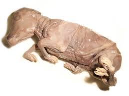
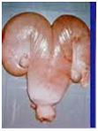
Maceration

Macerated fetus in the uterus
Maceration may occur at any stage of gestation and has been observed in all species. When the fertilized ovum or embryo succumbs to bacterial or viral infection or other disease or abnormality early in gestation it is usually absorbed in the uterus or a slight and often insignificant purulent uterine or vaginal discharge may be evident.
The embryo is seldom observed. The interval between estrrual periods may be prolonged if the embryo did not succumb until 20 to 50 days after conception. Early embryonic death and maceration are probably caused by a variety of miscellaneous organisms that may be found in the uterus, and are of common occurrence in cows affected with trichomoniasis or vibriosis.
In the cow fetal maceration that occurs after third month of gestation by which time fetal bones are fairly well developed, may be caused by similar wound ? infection bacterial agent. More commonly fetal emphysema and maceration follow fetal death and beginning abortion in the cow in which the cervix dilated but the fetus was not expelled due to a failure of the genital tract to dilate sufficiently or contract normally or because the fetus was dead and in an abnormal position and posture. In rare instances fetal emphysema and maceration may be associated with uterine torsion during gestation. Two factors of open cervix and a dead fetus at body temperature cause a rapid bacterial invasion of the fetus and membranes of organisms already present in the uterus or from the more caudal portions of the reproductive tract, fetal emphysema and maceration follow.
If the bovine fetus is beyond the third month of pregnancy and if the usual expulsive efforts are not observed or are unsuccessful, the fetus develops emphysema in 24 to 48 hours and in 3 to 4 days maceration begins.
Symptoms:
In long standing fetal maceration, the acute emphysematous stage has passed straining is seldom observed and the cervix is usually quite contracted. Generalized symptoms of elevated temperature and pulse and anorexia are usually not present.
There is often a history of chronic, fetid, mucopurulent discharge from the vulva over a period of several weeks or months.
There may be a history of a gradual drop in milk flow and loss of weight.
On rectal examination in the cow, fetal bones may be palpated in the uterus either floating in pus or crepitating with little pus around them. The uterine wall is thick and heavy and the cervix usually large and hard.
Treatment
The prognosis is poor. Treatment in the cow is difficult. If much pus is present, treatment as for Pyometra is indicated and possibly the bones may be passed through the cervix with the pus.
Source: http://univsul.edu.iq/Wenekan_KS/5018141612015_Lec,%206.pdf
Pyometra
Pyometra is characterised by a progressive accumulation of pus in the uterus and by the persistence of functional luteal tissue in the ovary.
In most cases, pyometra occurs as a sequel to chronic endometritis when, as noted above, as a result of inflammation, the uterus ceases to produce or release the endogenous luteolysin. The corpus luteum of dioestrus persists and, since the genital tract remains under the continuous influence of progesterone, the infection is not eliminated. Because the cervix remains fairly tightly closed the purulent exudate accumulates within the uterine lumen, although occasionally there is a slight purulent discharge. Occasional cases of pyometra occur in the presence of a luteal cyst.
The second main cause of pyometra is the death of the fetus, invasion of the uterus by A. Pyogenes and retention of the corpus luteum of pregnancy. This is a relatively infrequent cause of the condition.
Thus pyometra usually results from contamination of the uterus during dioestrus, such as occurs after insemination during the luteal phase. Venereal infection with organisms such as
Trichomonas fetus, which cause embryonic death, also causes pyometra.
Cows which suffer from pyometra show few or no signs of ill health; the main reason for them being examined is the absence of cyclical activity, or, perhaps, the presence of an intermittent vaginal discharge.
The uterine horns are enlarged and distended, quite often to an unequal degree, owing to incomplete involution of the previously gravid horn or to recent conceptual death.
Differentiation of pyometra from a normal pregnancy can sometimes be difficult, but there are a number of distinguishing points:
?The uterine wall is thicker than at pregnancy.
?The uterus has a more ‘doughy?and less vibrant feel.
?It is not possible to ‘slip?the allantochorion.
?In most cases of pyometra, no uterine caruncles can be palpated. However, when the infection occurred in a non-involuted uterus, involution of the caruncles is delayed and they may remain palpable for quite a long time.
?Transrectal ultrasonography will demonstrate the absence of a fetus and the presence of a ‘speckled?echotexture of the uterine contents compared with the black anechoic appearance of normal fetal fluids
Pyometra associated with T. foetus infection presents features which are different from those previously described. Uterine pus is, as a rule, much more copious and may attain a volume of many litres. It is generally more fluid and is greyishwhite or white. The uterus undergoes much greater distension. The mucus occupying the cervix is moist and slippery, rather than sticky and tenacious, and motile trichomonads can generally be found in it.
Treatment
The best treatment is the use of PGF2α or its analogues. They result in regression of the corpus luteum, dilatation of the cervix and expulsion of the purulent fluid, with oestrus occurring 3? days later.
If there is any doubt about the diagnosis of pyometra the cow should be left untreated and re-examined 2 weeks later for evidence of change Provided that the condition is not too longstanding and therapy is instituted quickly, there is a reasonable possibility that the cow will eventually conceive again.
However, long-standing cases are associated with more severe degeneration of the endometrium, reducing chances of reconception.
Repeat Breeder cow
To identify repeat breeder cows you need two things: good records and good heat detection. Given what has been said above and that on many farms the efficiency of oestrous detection is less than 60% (i.e. for every 10 cows potentially cycling only 6 are served) it can be seen that this is quite a need!
However if done well they allow the farmer or herdsperson to pick up cows that are cycling normally but not getting pregnant or most importantly those not fitting a normal pattern. Using this information these possible problem animals can be identified quickly, subjected to veterinary examination and a treatment protocol applied. This reduces the potential days open and so saves money.
The exact amount depends on farm circumstances but it more than pays for a regular veterinary visit on most farms.
Good heat detection needs an ability to recognise the signs of heat and time set aside to look carefully for these as in some cases they may not be very obvious. The 3-week calendar can be very useful in pinpointing likely candidates.
Other aids, such as beacons, tail paint pedometers and milk progesterones can also improve heat detection but there is still no really cost effective substitute for the astute observer apart from the bull. Even the latter can be overwhelmed if there are too many cows in season at one time.
Causes:
1. Early Embryonic death:
In this case the retention of CL of pregnancy, which has been terminated early, occurs. This is usually encountered during 90-120 days of gestation where the embryo is too small to be detected when aborted or it may be reabsorbed. This may be a characteristic feature in served cows coming into heat after a period longer than the normal oestrus cycle.
Treatment
? Improved nutrition.
? Suspect for Trichomoniasis and Vibriosis and treat them.
? Inj. receptal 11 days after insemination is known to improve embryo survival.
Anovulation:
Ovum not released from ovary. The animal has normal cycle, normal reproductive tract but fails to conceive.
Causes:
? Inadequate level or absence of L.H.
? Ovaro-bursal adhesion. It may be diagnosed by palpation of matured GF on the ovary more than 48hrs after the end of estrum.
Treatment:
? L.H preparations (HCG- human chorion Ganadrotrophin)- 3000 IU. I/V. when the animal is in heat.
? Inj. Receptal - 5 ml I/m.
? Improved feeding.
Delayed Ovulation:
Ovulation takes place 48-72 hours after the onset of oestrus but the spermatozoa would be dead by then.
Cause:
Due to low level of LH.
Treatment:
As for anovulation.
Preventing repeat breeder cows
Ensure you are serving cows at the correct time. This means that all staff should know the signs of heat. Milk progesterone testing is also useful; cows in a true heat will have very low progesterone.
Ensure insemination techniques are as good as possible. This is particularly important if you use DIY AI. Do not serve cows previously diagnosed as pregnant without doing a cow-side progesterone test to confirm it is has a low progesterone and is not pregnant. If the cow is pregnant AI may cause foetal loss.
Identify and treat cows with whites before starting to serve them.
Don’t start serving too soon after calving. Herds that start early have lower pregnancy rates to service and so more repeat breeder cows.
Minimise stress at service. For example, try and avoid serving around turnout or when you change the diet.
A comprehensive analysis and review of your breeding programme is time well spent in the battle to reduce repeat breeding. Get together with your vet and go through your herd records and present breeding protocols and see if there are not some areas where this important area could be improved.
Source: http://www.fwi.co.uk/livestock/repeat-breeding-syndrome.htm
Endometritis
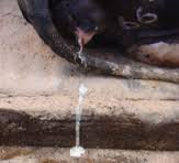
whitish/ mucopurulent vulval discharge from cow affected with endometritis
?span style="font:7.0pt "Times New Roman""> Endometritis is an infection of the uterine endometrium.
?span style="font:7.0pt "Times New Roman""> Cattle endometritis is a common condition that is known by the layman as 'whites'.
?span style="font:7.0pt "Times New Roman""> It occurs three weeks or more after calving and should not be confused with the more severe condition of metritis which occurs immediately post-partum.
?span style="font:7.0pt "Times New Roman""> The main consequence of endometritis is poor fertility. Therefore it has a major economic impact by increasing calving interval, services per conception and cull rates and by decreasing milk yield.
?span style="font:7.0pt "Times New Roman""> It is reported to have an incidence of between ten and fifteen percent in dairy herds (however it is very variable from herd to herd), with the total cost of £160 per case.
Etiology
?span style="font:7.0pt "Times New Roman""> The normally sterile uterus is contaminated by environmental microorganisms during parturition or immediately postpartum.
?span style="font:7.0pt "Times New Roman""> The main bacteria involved in endometritis is Trueperella pyogenes, however, numerous gram-negative anaerobes may also be involved.
?span style="font:7.0pt "Times New Roman""> The presence of this opportunist bacteria can delay return to service and cyclical activity, prevent fertilisation and cause early embryonic death by producing a hostile uterine environment. It is also reported that it increases incidence of ovarian cysts.
Signalment
?span style="font:7.0pt "Times New Roman""> Endometritis can occur in any cow post-partum however incidence is increased by the following predisposing factors:
?span style="font:7.0pt "Times New Roman""> Retained foetal membranes
?span style="font:7.0pt "Times New Roman""> Dystocia
?span style="font:7.0pt "Times New Roman""> Caesarian section or assisted calving
?span style="font:7.0pt "Times New Roman""> Induced parturition
?span style="font:7.0pt "Times New Roman""> Still Birth
?span style="font:7.0pt "Times New Roman""> Twins
?span style="font:7.0pt "Times New Roman""> Unhygienic calving environment - includes seasonal effect as indoor calving has higher endometritis rates
?span style="font:7.0pt "Times New Roman""> Ovarian inactivity
?span style="font:7.0pt "Times New Roman""> Parity
?span style="font:7.0pt "Times New Roman""> Concurrent disease and nutrition - fatty liver disease and hypocalcaemia are reported to increase endometritis rates.
?span style="font:7.0pt "Times New Roman""> Multiple defense mechanisms exist to prevent opportunistic infection of the uterus. The vulva and cervix provide a physical barrier to opportunist bacteria; resident bacteria in the vagina prevents the colonisation of harmful bacteria; involution and caruncle sloughing expel contaminants and tissue debris; a higher pH during oestrus prevent bacterial growth and various immunological mechanisms also act to prevent infection of the uterus. Following parturition endometritis may still occur, especially is one or more of the mentioned predisposing factors is present.
Clinical Signs
?span style="font:7.0pt "Times New Roman""> Mucopurulent vaginal discharge should be evident on vaginal exam 21 days or more post-calving. Discharge is relatively odourless (dependant on severity) and whites in colour, hence the name 'whites'.
?span style="font:7.0pt "Times New Roman""> The discharge should not be confused with lochial discharge or vaginitis.
?span style="font:7.0pt "Times New Roman""> Rectal palpation should reveal a poorly-involuted, oedematous uterus.
?span style="font:7.0pt "Times New Roman""> On an individual or herd level there may be a history of subfertility.
Diagnosis
?span style="font:7.0pt "Times New Roman""> Diagnosis should be based on the calving history and clinical signs following vaginal and rectal exam.
?span style="font:7.0pt "Times New Roman""> Vets may use a scoring system to categorise the colour and odour of the vaginal discharge which indicates how severe the infection is and whether treatment is necessary.
?span style="font:7.0pt "Times New Roman""> Measurements of the uterine and cervical diameter may be included in the scoring system. Definitive diagnosis can only be achieved by endometrial biopsy, however this is rarely indicated.
Treatment
?span style="font:7.0pt "Times New Roman""> Greater success is achieved with milder cases of endometritis. Treatments available include antibiotics, hormones and intrauterine antiseptics:
?span style="font:7.0pt "Times New Roman""> (1) Antibiotics
Various factors should be considered when selecting an antibiotic for the treatment of endometritis. Criteria for antibiotic:
appropriate efficacy in infected uterine environment
appropriate efficacy against the causal bacteria
no inhibition of natural uterine defense mechanisms
appropriate concentration and duration of action in the infected uterine lumen
little or no milk withdrawal period
cost effective
no detrimental effect on fertility
Generally, a broad spectrum antibiotic, active against Actinobaccillus pyogenes and gram-negative anaerobes should be used. Ideal antibiotics are cephalosporins and oxytetracycline as they match the majority of criteria listed above. Some resistance to oxytetracyclines is reported and additionally some formulations cause irritation to the endometrium, therefore intrauterine cephalosporin should be considered the most effective antibiotic treatment.
Sulphonamides, aminoglycosides, nitrofurazones and penecillins have decreased activity as a result of the uterine environment and bacteria present.
Metranidazole and chloramphenicol should not be used as they are banned from use in food-producing animals.
Some antibiotics are rapidly absorbed and distributed throughout the body, and in these cases it may be of more use to administer them parenterally.
(2) Hormones
Oestrogens: controversial treatment bases on the knowledge that the uterus is more resistant to infection during oestrus. It is not licensed in the EU for use in cattle.
Prostaglandins: PGF2a or analogues can be administered parenterally. They should be considered the treatment of choice if a corpus luteum is present. The administration of prostaglandins removes the inhibitory effect of progesterone on the uterus and induces oestrus, which both of these actions improve the uterine defense mechanisms. They may have an additional beneficial ecbolic effect, aiding clearance of the lumenal contents. There is no milk withdrawal period for prostaglandins, making them ideal for use in dairy cattle.
These are mainly used in chronic cases.
(3) Antiseptics
Chlorhexidine and metakresol sulphonic acid (Lotagen), povidone iodine antiseptic administered intrauterine are reported to be a effective alternative to antibiotic treatment, however few studies have been carried out to confirm this and detrimental effects on fertility are reported.
Prognosis
Prognosis is dependant on the severity of infection and the effectiveness of treatment. Greater success is seen with mild cases of endometritis.
Source: http://agridr.in/expert_system/cattlebuffalo/Obstetrics%20and%20Gynecological%20conditions.html
SILENT HEATS (UNOBSERVED AND SUBESTRUS)
Incidence of subestrus in cattle and buffaloes has a wide variation in the frequency (15 - 73%).
The intensity of heat signs in buffaloes is generally lower and also homosexual activity is not pronounced as in cows.
The intensity of expression of estrus is generally affected by housing, floor surface, yield, lameness and number of herd mates in estrus simultaneously.
Summer anestrous is an important condition is buffaloes contributing to infertility in buffaloes.
Heat stress has a pronounced effect on the estrus behavior of the female animals as heat stress reduces the length and intensity of estrus. Changes caused by heat stress reduce the likelihood of estrus detection.
Heat stress causes increased cortisol secretion and this hormone blocks estradiol-induced sexual behavior. Effect of heat stress on estrous behavior also includes actions independent of the pituitary-adrenal axis reducing the ovarian activity and follicular development. Heat stress causes a reduction in peripheral concentrations of estradiol at estrus. Moreover reduced physical activity is also probably an adaptive response that limits heat production and reduced estrous behavior.
Heat stress at the beginning on the day of ovulation reduces the diameter and volume of the dominant follicle of the estrous cycle. Secretion of the hormones regulating reproductive tract function is also altered by heat stress. These changes lead to variable changes in steroid hormone concentrations in peripheral blood. This reduced action of steroid hormones on reproductive tract tissue during heat stress eventually impairs uterine and oviductal functions.
Factors responsible for anestrus are environmental stress, endocrine (endogenous opioid peptide, lower insulin concentration, imbalance in hormones such as LH, FSH, prolactin, melatonin and thyroid), nutritional (negative energy balance, micro and macronutrient deficiency), managemental factors such as lactational stress, suckling etc. A lower level of body weight, total serum protein, blood hemoglobin, blood glucose, blood insulin, inorganic phosphorus, calcium, manganese, iodine, cobalt, copper, iron, cholesterol and vitamin A deficiency also causes anestrus. Anestrus may be diagnosed by per-rectal palpation of reproductive organs and frequent activity of reproductive cycles of buffaloes using ultrasonography.
Anoestrus
Anoestrus is the failure of a cow to cycle and display oestrus (heat). Anoestrus is normal before puberty, during pregnancy and for a short period after calving (postpartum anoestrus, PPA). All cows undergo varying degrees of PPA. On average, 20% of seasonal-calving cows are anoestrus at the start of mating, with extreme cases of over 50% of the herd also being reported.
Incidence of anestrus in buffaloes has been documented in various states as given below:
|
State |
Incidence (%) |
|
Haryana |
56-67 |
|
Punjab |
19.84 |
|
West Bengal |
50.84 |
|
UP |
69.40 |
|
Gujarat |
9.18 |
|
Tamilnadu |
9.09 |
|
Madhya Pradesh |
60.83 |
|
Andhra Pradesh |
30.67 |
|
Bihar |
44.60 |
|
Karnataka |
51.00 |
|
Andhra Pradesh |
56.36 |
Causes
The major causes of PPA in dairy cows are inadequate nutrition (negative energy balance, NEB), late-calving cows and uterine disease. NEB happens when energy intake is lower than output in milk production and maintenance. It is seen as a loss of body condition as body fat is mobilised to meet the energy demands of lactation.
Some degree of NEB is inevitable in nearly all dairy cows in the month after calving. Variation in BCS at calving, weather conditions, pasture availability and supplementary feeds from one year to the next will result in differences in the extent of NEB experienced, and this is reflected in submission rates early in the mating period.
Classification of anoestrus in cattle
Benefiting from the use of ultrasonography and growing knowledge of follicular dynamics in cattle Wiltbank et al. (2002) proposed the following classification of anovulatory status:
Anovulation with follicle growth up to the emergence stage
Cows exhibit very small follicles that are growing only to the emergence phase and do not proceed further.
Fairly common in cattle exposed to poor feeding conditions such as extensive pasture management in tropical zones.
The underlying factor is inadequate FSH stimulation
Anovulation with follicle growth up to deviation phase
Follicular growth takes place and proceeds through emergence and deviation but does not lead to ovulation. A frequently reported form of anoestrus. Commonly occurs in the post partum period in lactating dairy and suckled beef cows.
The characteristic signs of this condition are small ovaries, with the absence of a corpus luteum or ovulatory size follicles. The ovaries however show continuing follicular growth in a dynamic wave pattern up to the deviation phase.
The underlying physiological problem is an inhibitory effect of oestradiol on GnRH/LH pulses that does not allow the final growth or oestradiol production by the post deviation dominant follicle.
Pathogenesis of anovulatory status/anoestrus in cattle
Resumption of cyclic activity after calving is influenced by:
- nutrition
- body condition
- suckling
- lactation
- breed
- age
- uterine pathology
- debilitating disease
Fewer than 10% of cows fail to ovulate in most well-managed dairy herds by day 40 post partum. It is well established that poor nutritional status and Negative Energy Balance are responsible for the majority of anoestrus cases in both dairy and beef cattle.
Treatment of anovulatory/anoestrus conditions in cattle
Treatment is based on:
Improvement in energy status- optimal nutrition during the transition period and during early lactation.
Hormonal treatments- combined with increased energy supplementation or reduced suckling stimulus may also help to stimulate oestrus.
Anovulation with follicle growth up to the emergence stage
In these cases treatments with GnRH usually have no effect, as the follicles on the ovary do not have adequate LH-receptors to respond.
FSH/PMSG treatment combined with improved nutrition can increase follicle growth.
Start with PMSG (Folligon):
- 500-850 iu per cow of bos taurus type and crosses
- 300-350 iu per cow of bos indicus type
Once the follicular growth is increased GnRH (eg. Receptal; 2.5-5ml) can be used to stimulate maturation and ovulation of the dominant follicle.
Do not breed/AI in the first oestrus
Anovulation with follicle growth up to deviation phase

·
Ovsynch
protocol
- GnRH stimulation + induction of ovulation.
Click on thumbnail sketch for protocol.
· Modified Ovsynch protocol- second GnRH replaced by hCG (e.g. Chorulon; 1.500 IU).
Dairy cows can be successfully treated with simple or modified Ovsynch protocol as many anovulatory dairy cows have follicles of sufficient size and ovulatory capacity but do not have an LH surge.
Source: http://www.partners-in-reproduction.com/reproduction-cattle/treatment-anoestrus.asp
Culling of Dairy Animals
Dairymen should develop a checklist of culling reasons to use in their decision making process. The following list of 10 questions is one that could be used for each cow before deciding her future in the herd.
Is her yearly production 20 per cent or more below the herd's rolling average?
Is she a chronic mastitis case? Check this one closely, because a cow with chronic mastitis is producing below her capability and, in addition, could be spreading mastitis to other cows in the herd through the milking equipment.
Will she be dry four months or more? Long dry periods are costly to the dairyman and may indicate the cow has a problem of becoming pregnant, a trait not desired.
Is she a hard milker? Is her udder shape or teat structure such that she is a nuisance to milk?
Does she have a history of calving difficulties or post calving illnesses such as retained placenta, metritis, or milk fever? Cows that cause problems at calving time are not pleasant to have and are costly to keep in the herd.
Does she have an undesirable disposition? Is she a nervous cow or does she kick whenever her udder is touched? These are undesirable traits that should be noted along with production and calving problems.
Is she below the herd's average body type? Check body confirmation to see if it comes up to specifications for the herd.
Is she a timid cow? With the type of dry lot housing systems most dairymen have today, timid cows usually will not get the amount of feed required to be high producing animals.
Is she an old cow, and is the available barn space needed for freshening heifers? Fresh heifers usually have a higher genetic potential for milk production than older cows, especially if a progressive A.I. program is used in the herd.
A yes answer should probably be given to at least two questions before making the cow a strong candidate for culling. In many cases, though, one reason may be enough justification to base a culling decision.
Besides using a checklist in making culling decisions, other facts should be considered.
First-calf heifers:
In evaluating first calf heifers, consider the size of the heifers. Undersized heifers will probably produce less milk the first lactation because of their size. This situation is an indictment of the dairyman's heifer feeding program and not necessarily the producing potential of the heifer. So, among possible culls of equal performance, preference should probably be given to the younger, undersized heifer, especially if she improved during her lactation.
Stage of gestation:
Extremely long calving intervals can be costly. With other factors being equal, cows in mid-lactation with several months to go before freshening are better prospects for culling than cows that will freshen sooner and return to peak production sooner.
Mastitis:
Mastitis CMT (California Mastitis Test) scores, somatic cell counts should be checked carefully. Cows with CMT scores of 2, somatic cell counts of 1.2 million or greater, should be culled before other potential culls because they are potential sources of intramammary infections of other cows.
Age:
Other factors being equal, cull older cows before younger cows of comparable relative value. The genetic potential of younger animals should be greater than that of older animals, so keeping the younger cows in the herd longer should be a sound practice.
Past performance:
Given two cows with the same relative value and other factors equal, cull the cow first that has the lowest previous production records. Past performance can be suggestive of future potential. Management errors in the current lactation could adversely affect a cow's performance.
Introduction
In Goa, the dairy farmers mostly keep cows (indigenous and crossbred) along with few buffaloes for milk production. However, the genetic potential of the dairy animals is exploited fully, when they are fed with well balanced ration. Further, as feed cost is around 75% of the total cost of production, the success of the dairy farming depends not only on the quality of the ration, but also on the economy of the ration. Therefore, knowledge on the feeds and feeding of dairy animals is highly essential.
Cereals
.jpg) Grain form
Grain form
.jpg) Ground form
Ground form
Maize
(Crude Protein-10%; Ether extract-2.5%)
.jpg) Ground form
Ground form
Rice kani
(Crude Protein-10%; Ether extract-1.5-2.0%)
.jpg) Ground form
Ground form
Rice Polish
(Crude Protein-12-14%; Ether Extract-14-16%)
.jpg) Ground form
Ground form
Deolied Rice Polish
(Crude Protein-14-18%; Ether Extract-0.5-1.0%)
.jpg)
Brewers’s Dried Grains
(Crude Protein-25%; Ether Extract-5%)
.jpg)
Soybean Meal
(Crude Protein-48-50%; Ether Extract-0.5-1.0%)
.jpg)
Groundnut Cake
(Crude Protein-45%; Ether Extract-0.5-1.0%)
.jpg)
Mustard Seed Cake
(Crude Protein-35%; Ether Extract-7%)
.jpg)
Cottonseed Cake
(Crude Protein-25%; Ether Extract-7%)
.jpg)
Mineral Mixture
(Calcium-20%; Phosphorus-16%)
.jpg)
Common Salt
.jpg)
Cowpea Green Fodder
(Crude Protein-15-20%)
.jpg)
Maize Green Fodder
(Crude Protein-8-10%)
.jpg)
Napier Bajra Hybrid (CO-3) Green Fodder
(Crude Protein-8-10%)
.jpg)
Napier Bajra Hybrid (CO-4) Green Fodder
(Crude Protein-8-10%)
.jpg)
Paddy Straw
(Crude Protein-2.5-3.0%)
.jpg)
Dry Karad Grass
(Crude Protein-2.5-3.0%)
.jpg)
Green Karad Grass
(Crude Protein-5-6%)
.jpg)
Jowar Straw (Kadaba Kutti)
(Crude Protein-3.5-4.5%)
Bureau of Indian Standard (BIS) Specifications for Compounded Cattle Feed
Nutrients (%) |
Type-I |
Type-II |
Calf Starter |
Calf Grower |
Moisture (Max) |
11 |
11 |
10 |
10 |
Crude Protein (Min) |
22 |
20 |
23-26 |
22-25 |
Ether Extract (Min) |
3.0 |
2.5 |
4.0 |
4.0 |
Crude Fiber (Max) |
07 |
12 |
07 |
10 |
Acid Insoluble Ash (Max) |
3.0 |
4.0 |
2.5 |
3.5 |
Salt as NaCl (Max) |
2.0 |
2.0 |
--- |
|
Calcium (Min) |
0.5 |
0.5 |
--- |
|
Available Phosphorus |
0.5 |
0.5 |
--- |
|
Vit A (IU/kg) |
5000 |
5000 |
--- |
|
Calf starter is a balanced concentrate mixture, which is fed to the calves from 10th day of age to supplement the nutrients, when they are raised on limited milk intake.
Composition of calf starter
Ingredients |
Parts (kg/100 kg) |
Maize/ wheat shorts/ barley/ oats |
50 |
Groundnut cake/ soybean meal |
30 |
Skimmed milk powder |
07 |
Wheat bran/ rice bran |
10 |
Mineral mixture |
02 |
Common salt |
01 |
Vitamin A and D supplement (g/q) |
10 |
Available Phosphorus |
0.5 |
It is a mixture of different concentrate feed ingredients like cereal grains (maize, rice kani etc), cereal grain byproducts (rice polish, brewers?dried grains etc), oil cakes (soybean meal, groundnut cake, cotton seed cake etc), mineral mixture and common salt in different ratios as per the requirement.
Examples of Concentrate Mixture
Feed Ingredients |
Parts by Weight (kg) |
||||
Ex-1 |
Ex-2 |
Ex-3 |
Ex-4 |
Ex-5 |
|
Maize grain (ground) |
35 |
35 |
35 |
35 |
35 |
Soybean meal |
15 |
20 |
--- |
10 |
10 |
Groundnut cake |
15 |
--- |
24 |
--- |
15 |
Cotton seed cake |
--- |
12 |
24 |
27 |
14 |
Rice polish |
32 |
30 |
14 |
--- |
23 |
Brewers?dried grains |
--- |
--- |
--- |
25 |
--- |
Mineral mixture |
02 |
02 |
02 |
02 |
02 |
Common salt |
01 |
01 |
01 |
01 |
01 |
The concentrate mixture may be either in Mash Form or Pellet Form.
.jpg) Mash Form
Mash Form
.jpg) Pellet Form
Pellet Form
Feeding Schedule for calves, heifers, pregnant and lactating cows
Feeding schedule for calves (per day)
Age |
Whole milk (liters) |
Calf starter (kg) |
Legume (Cow pea) green fodder (kg) |
1-3 days |
3.0 (colostrums) |
--- |
--- |
4-15 days |
3.0 |
--- |
--- |
16-30 days |
3.5 |
Ad lib. |
Ad lib. |
1-2 months |
2.5 |
0.25 |
Ad lib. |
2-3 months |
2.0 |
0.50 |
2-3 |
3-4 months |
1.0 |
0.75 |
5-7 |
Feeding schedule for heifers (per day)
Age |
Diet (kg) |
|
Maize/ CO-3/ CO-4 Fodder |
Concentrate Mixture |
|
4-6 months |
6-7 |
1.5 |
6-12 months |
12-15 |
1.5 |
12-18 months |
20-25 |
1.0 |
18-30 months |
25-30 |
1.0 |
Pregnant (last quarter) |
30-35 |
2.0 |
Feeding schedule for pregnant animals 8-10 weeks before calving
Feeds |
Quantity (kg/ day) |
Maize/ CO-3/ CO-4 fodder |
30-35 |
Paddy straw/ Maize stover/ Kadaba Kutti/ Dry karad grass |
5 |
Concentrate mixture |
2-3 |
|
|
Feeding schedule for lactating animals
Rations for lactating animals (for cows producing up to 7 kg milk and buffaloes producing up to 5 kg milk per day)
Feeds |
Quantity (kg/ day) |
Maize/ CO-3/ CO-4 green fodder |
10-20 |
Paddy straw/ Maize stover/ Kadaba Kutti/ Dry karad grass |
7-8 |
Concentrate mixture |
3-4 |
|
|
Rations for lactating animals (for cows producing more than 7 kg milk and buffaloes producing more than 5 kg milk per day)
(i) If the dairy animal i.e. cows producing more than 7 kg milk and buffaloes producing more than 5 kg milk per day, then provide extra concentrate mixture @ 400g per kg increase in cow milk and 500 g per kg increase in buffalo milk.
(ii) If the dairy animal is pregnant, then besides the maintenance and production requirement, one kg concentrate mixture as pregnancy allowance should be offered extra only in the last three months of the pregnancy.
Feeding of high producing animals
The high yielding dairy animals need very special feeding strategies. Low quality roughages like straw or stovers should not be offered to high producing animas. The concentrate mixture of high yielders must contain energy and protein rich ingredients like soybean meal, roasted soybeans, by pass nutrients etc. Buffers like sodium bicarbonate, magnesium oxide and vitamin mixture should be added in the diet for better utilization of the feedstuffs. The composition of concentrate mixture for high yielders is presented in Table 11.
Concentrate mixture for high yielding animals
Ingredients |
kg |
Maize |
40 |
Mustard cake |
20 |
Soybean meal |
10 |
Roasted soybean |
05 |
Rice bran |
15 |
Wheat bran/ Deoiled rice bran |
07 |
Mineral and vitamin mixture |
02 |
Common salt |
01 |
(i) The green fodder must be chopped before feeding to the dairy cows for better utilization.
(ii) If possible, soak the concentrate mixture in water for 6-8 hours and then feed to the animals.
(iii) The required concentrate mixture, green fodder and straw may be offered either separately or mixing together as total mixed ration (TMR).
(iv) The total ration to be offered daily should be divided and offered twice (morning and afternoon) for better utilization.
(v) Do not feed calcium rich feed ingredients or mineral mixture to the dairy cows 15 days before parturition, as high calcium intake during this period increases the chances of milk fever.
(vi) Provide clean fresh water free of choice to the dairy cows. For easy accessibility, a cemented water tank should be constructed near to the cow shed and the tank should be painted with lime at frequent intervals to make the water clean.
Cattle are susceptible to a wide variety of infectious diseases. Susceptibility has increased with the introduction of exotic germplasm and climate changes. The important infectious diseases of the cattle and its control are:
Foot and mouth disease:
It is a highly infectious and contagious viral disease of all cloven footed domesticated and wild animals. The infection imposes a high spread during the cooler season when the air remains in damp condition.
Vaccination is the only means to control the disease.Vaccine |
Age |
Dose |
Booster |
Interval |
Season |
FMD |
6-8 wks |
10 ml s/c |
6, 9 or 12 months |
Annual |
Preferably November, December |
|
|
Black Quarter
Black Quarter is one of the deadly bacterial diseases of cattle. The disease spreads rapidly after heavy rainfall by contamination of soil with spores of the organism. Areas where previous death occurs from clostridium infection, have a higher incidence or risk of disease because of increased environmental contamination. Treatment of the disease with specific antibiotic is useful if the diagnosis of the disease can be made at the early stage.
The disease can be controlled by regular vaccination.Vaccine |
Age |
Dose |
Booster |
Interval |
Season |
Polyvalent B.Q. Vaccine |
All age |
5 ml s/c |
6 months |
Annual |
Before onset of monsoon, all season in endemic areas |
|
|
Haemorrhagic septicaemia:
Haemorrhagic septicaemia, as the name implies is a poisoning of the blood accompanied by internal haemorrhages, is an acute septicaemic disease caused by a bacterium, Pasteurella multocida. The incidence of H.S. is throughout the year. The outbreak is not only in the period of high humidity or monsoon but also in winter season. Highest number of outbreaks during high humid and rainy season as compare to winter season. Although antibiotics are available for the treatment, there may not be any time for treamtn in most cases.
The disease may be controlled by regular vaccination.Vaccine |
Age |
Dose |
Booster |
Interval |
Season |
H.S. adjuvant vaccine |
All ages |
3 ml i/m |
6 months |
Annual |
Preferably in May/June |
|
|
Brucellosis:
Brucellosis is one of the major zoonotic diseases and causes great economic losses due to loss of milk production, abortion at late pregnancy and high rate of infertility in females and varying degree of sterility in males. There is no seasonal variation for occurrence of the disease. Regular screening of serum samples for detection of the disease is required. Incidence of brucellosis can be reduced by elimination of infected bull, isolation and treatment of positive reactors and calfhood vaccination.
Brucella is one of the world’s major zoonotic pathogens, and is responsible for enormous economic losses as well as considerable human morbidity in endemic areas.
Pathogenesis :
Brucellosis is a sub-acute or chronic disease which may affect many species of animals. In cattle, sheep, goats, other ruminants and pigs the initial phase following infection is often not apparent. Brucella has predilection for the pregnant uterus, udder, testicle and accessory male glands, lymph nodes, joint capsules and bursae. In sexually mature animals the infection localizes in the reproductive system and typically produces placentitis followed by abortion in the pregnant female, usually during the last third of pregnancy, and epididymitis and orchitis in the male. Lesions consist of necrotic placentitis and interstitial mastitis in pregnant cows, and fibrinous pleuritis with interstitial pneumonia in aborted fetuses and newborn calves. The recently recognized types associated with marine animals may also have the capacity to cause human disease.
How to diagnose?Diagnosis can only be confirmed by laboratory tests. The isolation and identification of Brucella offers a definitive diagnosis of brucellosis and may be useful for epidemiological purposes and to monitor the progress of a vaccination programme in animals. The RBPT, tube agglutination and ELISA procedures are recommended. Culture of Brucella from abortion material, milk or tissues collected at autopsy provides a definitive diagnosis.
Facilities for diagnosis of brucellosis are available at ICAR-CCARI, Goa.
Differential diagnosis :Brucella abortions should be differentiated from septic abortion, abortion from other infectious agents such as listeriosis, Q fever, leptospirosis, campylobacteriasis, trichomonads, fungi, epizzotic viral abortion and infectious bovine rhinotracheitis.
Impact of the problem:Expansion of animal industries and urbanization, and the lack of hygienic measures in animal husbandry and in food handling partly account for brucellosis remaining a public health hazard. Expansion of international travel which stimulates the taste for exotic dairy goods such as fresh cheeses which may be contaminated, and the importation of such foods into Brucella-free regions, also contribute to the ever-increasing concern over human brucellosis. Sexual transmission and/or artificial insemination are also important.
The disease is of zoonotic importance.
Treatment:Treatment for brucellosis in animals is neither advisable nor practicable.
In humans :
Treatment of uncomplicated cases in adults and children eight years of age and older: doxycycline 100 mg twice a day for six weeks + streptomycin 1 g daily for two to three weeks.
OR
?Doxycycline 100 mg twice a day for six weeks + rifampicin 600?00 mg daily for six weeks.
Control and Prevention :?Animal brucellosis is best prevented by careful herd management and hygiene.
?Vaccination is useful for prevention and control of infection.
?B. abortus strains 19 and RB 51 are recommended for prevention of bovine brucellosis.
?B. melitensis Rev 1 is recommended for prevention of B. melitensis infection in sheep and goats.
?Control and prevention schemes require effective collaboration between all sections of the community.
?Education and information programmes are essential to ensure cooperation at all levels in the community.
?Effective preventive measures and control of animal movements are essential.
Listeriosis :
Listeriosis is an important bacterial infection, which occurs in an array of animals including humans. It arises mainly from the ingestion of contaminated food. Listeriosis leads to septicaemia, abortion, stillbirth and menigoencephalitis.
Pathogenesis :Ingestion of Listeria from food --> Intestine --> Mean incubation period: 31 days --> L.monocytogenes crosses mucosal barrier --> Bloodstream --> Hematogenous dissemination --> Brain, placenta and other sites.
How to diagnose?The disease can be tentatively diagnosed on the basis of clinical signs and symptoms. Its confirmation is achieved by isolating the pathogen from clinical specimens, such as blood, cerebrospinal fluid (CSF) and the muconium of the newborn or foetus in abortion cases, and from faeces, vomitus, food stuffs/animal feed and/or vaginal secretions of infected individuals.
Differential diagnosis :Listeriosis has been difficult to diagnose as it can easily be confused with the nervous form of acetonaemia in cattle, early cases of pregnancy toxaemia in sheep, rabies and encephalitis of viral origin. Listerial abortion must be differetaited from other causes of abortion like brucellosis, leptospirosis and trichomonads.
Impact of the problem :The spectrum of the disease in animals is broad ranging from asymptomatic infection and carriage to uncommon cutaneous lesions or various focal infections such as conjunctivitis, urethritis, endocarditis, and severe disturbance of the gait, followed by death. Abortions and perinatal deaths are common in cattle and sheep. L. monocytogenes is a well-recognized cause of mastitis, abortion, repeat breeding, infertility, encephalitis and septicaemia in cattle.
Listeriosis is an important bacterial zoonosis which occurs in a variety of animals including humans, and arises mainly from the ingestion of contaminated food and water. Listeriosis is a serious invasive disease which leads to septicaemia, abortion, stillbirth, meningitis and meningoencephalitis, especially in immunocompromised individuals and in persons in contact with animals.
The prevalence of listeriosis is decidedly more among the individuals with immunocompromised status, young age groups, elderly people and persons who are immunosuppressed due to medication and/or illness.
Treatment:Ampicillin, amoxicillin, tetracyclines, chloramphenicol, trimethoprim and sulphamethoxazole are recommended. Ampicillin is the drug of choice in cases of encephalitis. Ampicillin along with gentamicin are recommended for prolonged treatment regimes.
Control and Prevention?Culling infected animals should be advocated as they secrete the organisms in secretions and excretions, especially in the cases of mastitis.
?Care in the use and preparation of silage is important as the pathogen grows luxuriantly at a pH greater than 5.
?So far no vaccine is available against listeriosis.
?Listeriosis can be prevented in humans by taking care during handling of the abortion cases in both humans and in animals, avoiding consumption of contaminated food-stuffs and avoiding cross-infections especially among infants in hospitals.
Mastitis :
Mastitis or inflammation of mammary gland or udder is an economically important condition in milking animals. Mastitis is a disease that affects a large number of dairy cattle throughout the world. It may affect one or all quarters of the udder. It is manifested in the form of swelling of the affected quarters, change in milk colour and composition.
PathogenesisDevelopment of mastitis is complex and involves three stages: invasion, infection and inflammation. The factors which contribute to the causation of mastitis include-
v Contamination with milker's hands, contaminated floors, utensils and clothes.
v Presence of high population density of the causative bacteria in milking shed.
v Damage to the teat sphincter.
v Udder infected with FMD, Pox virus.
v Physical trauma that may accelerate the growth of these causative bacteria.
v Sawdust and shaving used as bedding, which are harboring E. coli and Klebsiella pneumoniae.
v Edema and congestion of udder during parturition.
v Poorly designed housing, uneven faulty surface.
v Dirty milking machine.
How to diagnose?
Clinical signs, palpation of udder, monitoring the milk consistency may give clue about causation of mastitis. Diagnosis of mastitis is based on bacteriological and cytological methods of examination. Subclinical cases may go unnoticed and therefore testing of milk with California mastitis test (CMT) or any other spot test is necessary. Bacteriological examination of CMT positive samples should be carried out.
Impact of the problemIf one quarter is affected by mastitis, 25% milk yield is lost permanently. It may also spread to other quarters, hence it causes major economic losses to the farmer. High yielding animals are more prone to this condition, if not milked properly. The milk from infected udders contributes to high microbial counts of milk, which in turn is not suitable for preparation of milk products. Mastitic organisms are also pathogenic for human beings. Mastitis control is prerequisite to any of the clean milk production programmes.
Treatmenta. Udder infusion:. Disposable tubes with water soluble ointment base need to be used. Emptying of udder is essential before infusion. For animal with clinical mastitis manual removal of congestion in the gland and inflammatory debris from the duct system should be done.
b. Parental treatment is advisable. Before antibiotic therapy, the sensitivity of the pathogens to particular antibiotics should be carried out.
c. The use of preparations containing disinfecting ingredients and added emollients is effective which promote the healing of teat lesions and prevent new infections.
Iodophors are important teat dips. Application of preparations containing 3-5000 mg. available iodine per kg, depending on the glycerin concentration, leads to reduction in bacterial numbers.
d. Culling of cows with therapy resistant udder diseases.
e. If machine milking is practiced then rinsing of the teat cups and dipping them in the disinfectant wash water between cows, by dipping a pair of each cluster in turn or hot water (170°-180°) for 10 sec. is effective.
In case of dry cows, infusion at the time of the last milking or at the beginning or end of the dry period is recommended.
Control and Prevention?Mastitis control also entails a good understanding of the factors that encourage its incidence and the microorganisms that cause it.
?Mastitis control must be concentrated on the prevention aspects, which depends mainly on the whole hygienic management and absence of stress conditions.
?Estrogenic compound in foods should be reduced. Young animals should be milked before older. Newly introduced animal should milked separately.
?Specific control measures need to be taken according to the respective cause and the extent of losses. Specific control measures include -
1. Correction of milking technique.
2. Teat disinfection (e.g. teat dipping) following milking.
3. Antibiotic treatment at drying off.
4. Culling of animals with therapy - resistant mastitis.